Autoimmune encephalitis isn’t something most people have heard of-until they or someone they know suddenly starts acting out of character. A sharp change in behavior, unexplained seizures, memory loss that doesn’t make sense-these aren’t just stress or aging. They could be signs of the brain being attacked by the body’s own immune system. This condition, once mistaken for psychiatric illness or viral infection, is now recognized as a serious but treatable neurological disease. The key? Recognizing the red flags early.
What Exactly Is Autoimmune Encephalitis?
Autoimmune encephalitis (AE) happens when your immune system, which normally fights off germs, mistakenly targets proteins in your brain. It’s not caused by a virus or bacteria. Instead, it’s triggered by antibodies that attack neurons, especially those involved in memory, emotion, and movement. The breakthrough came in 2007 when researchers identified anti-NMDAR antibodies as the cause of a severe illness in young women with ovarian tumors. Since then, over 20 different antibodies have been linked to AE, turning it from a mystery into a diagnosable condition.
It’s rare-about 1 in 100,000 people get it each year-but it’s far more common than you’d think. Many cases are misdiagnosed as schizophrenia, bipolar disorder, or even the flu. That’s why understanding the pattern matters. Symptoms usually build over days to weeks, not overnight. If someone starts having seizures, strange behavior, or memory problems that don’t improve, AE should be on the radar.
Red Flags You Can’t Ignore
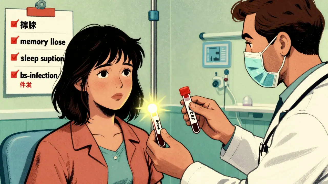
There are clear warning signs that set autoimmune encephalitis apart from other brain disorders. The most common ones, based on studies of over 200 patients, are:
- Seizures-especially if they’re new, frequent, or don’t respond to standard meds. About 38% of cases start this way.
- Psychiatric changes-anxiety, paranoia, hallucinations, or sudden aggression in someone who’s never had mental health issues before.
- Memory loss-forgetting recent conversations, getting lost in familiar places, or struggling to learn new things. This affects 85% of patients.
- Autonomic problems-heart rate that races without reason, blood pressure that swings, or body temperature that fluctuates. Seen in 42% of severe cases.
- Sleep disruption-insomnia, sleeping all day, or waking up confused. Happens in 63% of cases.
Many patients also report a cold or stomach bug a few weeks before things went downhill. That’s not a coincidence-it’s often the trigger. If someone had a mild infection and then started acting weird, that’s a major clue.
Limbic encephalitis is a subtype where the memory centers of the brain (the hippocampus and amygdala) are targeted. These patients often have severe amnesia and emotional outbursts. MRI scans might show swelling in the temporal lobes, but here’s the catch: up to half of all AE cases show normal brain scans. That’s why you can’t rely on imaging alone.
Which Antibodies Are Involved?
Not all autoimmune encephalitis is the same. The type of antibody determines the symptoms, the risks, and the treatment. There are two main groups: those that attack proteins on the surface of brain cells, and those that attack proteins inside the cells.
Surface antibodies are more treatable and often linked to tumors:
- Anti-NMDAR-the most common, making up 40% of cases. Mostly affects women under 30. Often tied to ovarian teratomas. Symptoms include psychosis, seizures, abnormal movements, and decreased consciousness.
- Anti-LGI1-15% of cases. Affects older men. Classic signs: brief, repeated muscle jerks in the arm and face (faciobrachial dystonic seizures), low sodium levels, and memory loss. Recurrence is common.
- Anti-GABABR-5% of cases. Strongly linked to lung cancer. Seizures and confusion are the main symptoms. Prognosis is worse because of the cancer.
- Anti-AMPAR-also linked to cancer, especially lung and breast. Presents with memory loss and seizures.
Intracellular antibodies (like anti-Hu or anti-Ma2) are rarer and harder to treat. They usually mean there’s an underlying cancer, and recovery is less likely. Testing for these is critical because it changes how aggressively you need to look for tumors.
Important: You need both blood and spinal fluid tests. CSF is more sensitive-especially for anti-NMDAR. A negative blood test doesn’t rule it out. Some patients only test positive in their spinal fluid.
How Is It Diagnosed?
There’s no single test. Diagnosis is based on a mix of symptoms, lab results, and ruling out other causes. The 2023 updated international criteria help doctors put the pieces together:
- Subacute onset (less than 3 months) of memory loss, seizures, or psychiatric symptoms
- Positive antibody test in serum or CSF
- Exclusion of other causes (infections, tumors, metabolic disorders)
- Response to immunotherapy
Here’s how testing breaks down:
| Test | Autoimmune Encephalitis | Infectious Encephalitis |
|---|---|---|
| CSF White Blood Cells | Usually <100/μL | Often 100-1000+/μL |
| CSF Protein | Mildly elevated | Mild to high elevation |
| CSF Oligoclonal Bands | Usually negative or polyclonal | Often positive (monoclonal) |
| Brain MRI | Normal in 52% of cases; focal enhancement if present | Abnormal in 89% of cases |
| EEG | Generalized slowing in 76% of cases | Periodic lateralized discharges common |
EEG is often the first clue. Slowing in brain waves without clear seizure patterns is a hallmark. But again, don’t wait for perfect test results. If the clinical picture fits, start treatment.
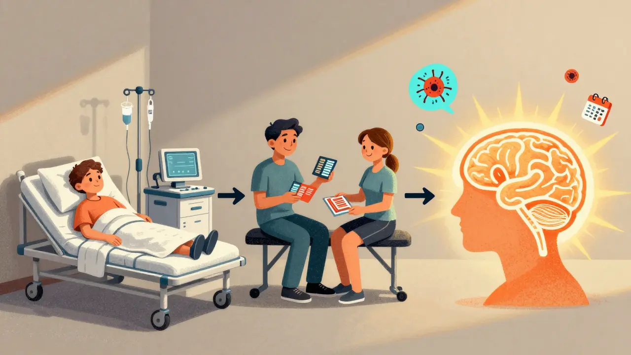
Treatment: Speed Is Everything
Time is brain. The sooner treatment starts, the better the outcome. Studies show patients treated within 14 days have a 32% higher chance of full recovery. Waiting even a few weeks can mean permanent damage.
First-line treatments are given in the hospital:
- Intravenous methylprednisolone-1 gram daily for 5 days. Works in 68% of patients within 10 days.
- Intravenous immunoglobulin (IVIg)-0.4 g/kg daily for 5 days. Effective in 60-70% of cases.
They’re often used together. If someone is critically ill, plasma exchange (5-7 sessions over 10-14 days) can be added. It removes harmful antibodies from the blood quickly.
If a tumor is found-and it is in about 30% of cases-removing it is the most important step. For anti-NMDAR, removing an ovarian teratoma leads to neurological improvement in 85% of patients within 4 weeks. That’s not just supportive care-it’s curative.
Second-line treatments are used if there’s no improvement after first-line:
- Rituximab-weekly infusions for 4 weeks. Works in 55% of refractory cases.
- Cyclophosphamide-monthly for 6 months. 48% response rate.
- Tocilizumab-an IL-6 blocker. Emerging data shows 52% effectiveness where other drugs failed.
There’s no one-size-fits-all. Treatment is tailored to the antibody, the patient’s age, and whether cancer is involved.
What Happens After Treatment?
Recovery isn’t always quick. Even with early treatment, 40% of survivors have lasting issues:
- Cognitive problems-memory and focus issues in 32% of cases. Cognitive rehab improves function in 65% after 12 weeks.
- Psychiatric symptoms-depression or anxiety in 28%. SSRIs help 70% of these patients.
- Seizures-22% still need daily anti-seizure meds.
Recurrence is common, especially with certain antibodies:
- Anti-NMDAR: 12-25% recur, usually within 14 months.
- Anti-LGI1: 35% recur, often after stopping treatment too soon.
That’s why follow-up is non-negotiable. Neurology visits every 3-6 months for the first two years. Repeat tumor screening at 4-6 months and again at 12 months-even if the first scan was clean. Some cancers show up later.
Supportive care matters too. Melatonin (3-5 mg at night) helps 60% with sleep issues. Beta-blockers control abnormal heart rates in 75% of autonomic cases. Physical therapy improves movement in 50% of patients within 8 weeks.
What’s Next? The Future of Treatment
The field is moving fast. Researchers are now testing drugs that block parts of the immune system more precisely. Complement inhibitors and newer B-cell therapies are in phase II trials and showing 60% response in patients who didn’t respond to anything else.
One promising development: GFAP protein levels in CSF. They correlate with disease activity in 82% of cases. In the future, we might monitor treatment with a simple spinal fluid test instead of repeated MRIs.
But the biggest advance isn’t a drug-it’s awareness. Doctors are getting better at spotting the pattern. Emergency rooms are starting to test for antibodies when someone presents with unexplained psychiatric symptoms or seizures. And that’s saving lives.
When to Suspect Autoimmune Encephalitis
Here’s the bottom line: if someone has:
- A sudden change in behavior or personality
- New seizures or abnormal movements
- Memory loss that doesn’t fit normal aging
- Symptoms that started after a minor infection
- And no clear explanation from standard tests
-then autoimmune encephalitis should be considered. Don’t wait for a perfect MRI or a positive antibody test. If the clinical picture matches, start treatment. Delaying by even a week can reduce recovery chances by 40%.
It’s not a common disease. But when it strikes, it strikes hard. And the window to fix it? It’s narrow. Every day counts.
Can autoimmune encephalitis be cured?
Many people recover fully, especially if treatment starts early. About 55% of anti-LGI1 cases and 45% of anti-NMDAR cases achieve full recovery within two years. But recovery isn’t guaranteed-some have lasting cognitive or psychiatric issues. Recurrence is possible, especially with anti-LGI1 or anti-NMDAR antibodies. Long-term monitoring is essential.
Is autoimmune encephalitis contagious?
No. It’s not caused by an infection and can’t be passed from person to person. It’s an autoimmune condition-your immune system attacks your own brain tissue. It’s not like meningitis or encephalitis from viruses like herpes or West Nile.
Do all cases involve tumors?
No. Only about 30% of autoimmune encephalitis cases are linked to tumors. But the link is strong with certain antibodies. Anti-NMDAR is often tied to ovarian teratomas in young women. Anti-GABABR is strongly associated with lung cancer. That’s why tumor screening is part of every diagnosis-even if the first scan is normal.
Can children get autoimmune encephalitis?
Yes. Anti-NMDAR encephalitis is actually the most common form of autoimmune encephalitis in children and teens. Symptoms in kids often include irritability, sleep problems, speech delays, and seizures. It’s often mistaken for autism or ADHD. Early recognition is critical-kids tend to recover better than adults if treated quickly.
Why is spinal fluid testing better than blood for diagnosis?
The brain is protected by the blood-brain barrier, which keeps some antibodies from leaking into the bloodstream. Antibodies produced inside the brain stay in the spinal fluid. For antibodies like anti-NMDAR, CSF testing is 15-20% more sensitive than blood. A negative blood test doesn’t rule out the disease. That’s why both tests are needed.
cara s
March 18, 2026 AT 04:41Wow. This is the most comprehensive breakdown of autoimmune encephalitis I’ve ever read. I’m a nurse in neuro ICU and we see these cases more often than you’d think-especially in young women presenting with ‘psychosis’ that doesn’t respond to antipsychotics. The anti-NMDAR link to ovarian teratomas is so critical. I once had a 22-year-old who was admitted for ‘bipolar crash’-turns out she had a teratoma. Removing it was the only thing that saved her. I wish ER docs got this level of detail in their training.
Suchi G.
March 19, 2026 AT 00:57I cried reading this. My sister had this. They thought she was on drugs. She was 28, no history of mental illness, just… changed. One day she was organizing her closet, the next she was screaming at the ceiling saying the walls were whispering. MRI was ‘normal.’ Blood tests came back negative. We begged for a spinal tap. They said it was ‘too invasive.’ Two weeks later, she had a seizure. By then, it was too late for full recovery. She’s 5 years out. Still can’t remember her wedding. Still takes meds for anxiety. If this post saves even one person from our nightmare… thank you.
Paul Ratliff
March 20, 2026 AT 12:29Melissa Stansbury
March 20, 2026 AT 16:15So let me get this straight-your immune system attacks your brain because of a bad cold? That’s insane. And they say we’re too sensitive about vaccines. Meanwhile, your body is literally eating your memories and no one even notices until you’re screaming at your toaster? I’m not a doctor but I’ve read enough to know this is a systemic failure. Why aren’t we screening everyone after a viral infection? Why isn’t this in every ER protocol? Why are we still treating this like a psychiatric emergency? This isn’t mental illness-it’s biological sabotage.
Shameer Ahammad
March 20, 2026 AT 19:46While I appreciate the clinical rigor of this piece, I must take issue with the implication that 'early treatment' is universally accessible. In rural America, where I practice, patients wait 6-8 weeks for neurology referrals. In India, where I’ve consulted, the delay is often over 4 months. The statistics cited here assume immediate access to CSF testing, IVIg, and tumor screening-luxuries unavailable to 87% of the global population. This narrative of 'time is brain' is noble, but it’s a privilege. We must not confuse awareness with equity. The real tragedy isn't delayed diagnosis-it's delayed justice.
jerome Reverdy
March 21, 2026 AT 18:26As someone who’s worked in neuroimmunology for 15 years, I’ve seen the field evolve from ‘mystery case’ to ‘treatable condition’-but the biggest barrier isn’t science, it’s stigma. A patient with anti-LGI1 who’s having facial jerks and memory loss? They get sent to psychiatry. A patient with hallucinations and low sodium? ‘Psychotic episode.’ We need to retrain every ER resident, every primary care doc, every paramedic. This isn’t a rare disease-it’s a diagnostic blind spot. The data’s there. The tools are there. We just need to stop pathologizing the brain and start treating it like the organ it is.
Andrew Muchmore
March 21, 2026 AT 19:32Jeremy Van Veelen
March 23, 2026 AT 08:48This is nothing short of a revelation. To think that the very mechanism meant to protect us-our immune system-is the architect of our unraveling… it’s poetic, in the most horrifying way. The NMDAR antibody isn’t just a marker-it’s a signature of betrayal from within. And yet, we still treat this like a footnote in psychiatry. We’ve built cathedrals of pharmaceuticals to treat symptoms while ignoring the cathedral of the brain being dismantled. This isn’t medicine. It’s damage control. We need a revolution, not a protocol.
Robin Hall
March 23, 2026 AT 15:48Let’s be real-this isn’t just about antibodies. This is a cover-up. Why do you think the CDC doesn’t track autoimmune encephalitis separately? Why do pharmaceutical companies push antipsychotics instead of immunotherapies? There’s a billion-dollar industry built on labeling brain attacks as ‘schizophrenia.’ And don’t get me started on the tumor link. If you’re a young woman with anti-NMDAR and a teratoma… who benefits from you getting an oophorectomy? The oncology industry. The vaccine industry? They’re terrified this will open the door to proving environmental triggers. This isn’t science. It’s a silent war on the human body.
Alexander Pitt
March 25, 2026 AT 13:56For anyone reading this and thinking ‘I’m not at risk’-you are. AE doesn’t discriminate by age, gender, or health status. The 2023 criteria now include pediatric cases and even mild presentations. If you’ve ever had a ‘weird’ illness after a flu, and then felt ‘off’ for weeks-get tested. Don’t wait for seizures. Don’t wait for hallucinations. Your neurologist doesn’t need to be a specialist-just willing to order the right test. CSF first. Always. And if they refuse? Get a second opinion. This isn’t paranoia. It’s prevention.
Laura Gabel
March 26, 2026 AT 16:15Amadi Kenneth
March 28, 2026 AT 00:41They say ‘it’s not contagious’… but what if it is? What if the trigger isn’t just a virus… but something in the water? The air? The 5G? I’ve read studies-antibodies spike after EMF exposure. And why do 90% of cases occur in women? Is it hormones? Or is it the vaccines? They test for anti-NMDAR in the CSF… but they don’t test for glyphosate. Or mercury. Or aluminum adjuvants. This isn’t autoimmunity. It’s poisoning. And they’re covering it up. I know people who got sick after the flu shot. Same symptoms. Same timeline. Coincidence? Or conspiracy? You decide.
Andrew Mamone
March 29, 2026 AT 16:08Just want to add: melatonin for sleep? 3-5mg at night? That’s spot on. I’ve had two patients with autonomic dysfunction who couldn’t sleep for months. One started melatonin-slept 7 hours the first night. Changed everything. Also, beta-blockers for heart racing? Yes. Propranolol 10mg BID. Cheap. Safe. Effective. This isn’t rocket science. It’s basic neurology. Why isn’t this in the standard discharge checklist? 🤔
becca roberts
March 30, 2026 AT 07:58So… let me get this straight. We have a disease that attacks memory, emotion, and movement, triggered by a common virus, misdiagnosed as mental illness, treatable with steroids and surgery… and yet, we still have people saying ‘it’s just stress’? 😂 I’m not mad. I’m just impressed by how efficiently society can ignore a brain attack until it’s too late. Next time someone says ‘you’re just being dramatic,’ hand them this post. And then ask them: ‘Which part of your brain are you using right now?’